Penyakit seliak
Penyakit seliak ialah gangguan autoimun jangka panjang yang terutamanya memberi kesan kepada usus kecil. Gejala-gejala klasik termasuk masalah perut usus seperti cirit-birit kronik, kembung abdomen, malabsorpsi, kehilangan selera makan dan di kalangan kanak-kanak tidak dapat membesar secara normal.[5] Ini sering bermula antara enam bulan dan dua tahun.[5] Gejala tidak klasik adalah lebih biasa, terutama pada orang yang lebih tua daripada dua tahun.[6][7][8][9] Mungkin terdapat simptom gastrousus yang ringan atau tidak, pelbagai gejala yang melibatkan mana-mana bahagian badan atau tidak ada gejala yang jelas.[5]
| Penyakit seliak | |
|---|---|
Nama lain | Spesis seliak, semulajadi nontropi, semulajadi endemik, enteropati gluten |
 | |
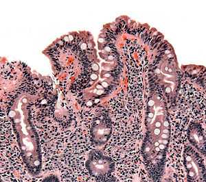
| Biopsi usus kecil menunjukkan penyakit seliak yang ditunjukkan oleh villi yang mengontot, hipertropi krip, dan penyusupan krip limposit | |
| Pengkhususan | Gastroenterologi, perubatan dalaman |
| Gejala | Tiada atau tidak spesifik, proses menggelembung perut, cirit-birit, sembelit, malabsorpsi, penurunan berat badan, dermatitis herpetiformis[1] |
| Kerumitan | Anemia kekurangan zat besi, osteoporosis, ketidaksuburan, barah, masalah neurologi, penyakit autoimun lain[2][3] |
Permulaan biasa | Sebarang usia |
| Tempoh | Sepanjang hayat |
| Punca | Tindak balas kepada gluten |
| Sejarah keluarga, ujian antibodi darah, biopsi usus, ujian genetik, tindak balas kepada penarikan gluten | |
| Penyakit usus radang, parasit usus, sindrom rengsa usus, sistik fibrosis[4] | |
| Rawatan | Diet/pemakanan bebas gluten |
| Kekerapan | ~1 dalam 135 |
| sunting | |
Penyakit seliak pertama kali digambarkan pada zaman kanak-kanak;[6][10] Walau bagaimanapun, ia boleh berkembang pada bila-bila masa.[5][6] Ia dikaitkan dengan penyakit autoimun lain, seperti diabetis melitus jenis 1 dan tiroiditis, antara lain.[10]
Tanda dan gejala sunting
Pucat, tinja atau kotoran yang berminyak (steatorrhoea) dan penurunan berat badan atau sulit untuk menambahkan berat badan merupakan gejala klasik penyakit ini. Ada juga kes seliak yang bergejala tidak terlihat yang pada organ selain dari usus itu sendiri,[11] malah penyakit celiac boleh dideritai tanpa sebarang gejala lazim[12] - keadaan ini dilihat pada 43% kasus diperhatikan dalam kalangan anak-anak.[13]
Banyak individu dewasa yang berpenyakit ini hanya mengalami kelelahan atau anemia.[14]> Kebanyakan mereka yang tidak diagnosis pada asalnya menyangka diri tidak bergejala kerana keterbiasaan mereka mengalami keadaan yang seolah-olah normal atau biasa; justeru mereka dapat memahami keadaan sebenar serta gejala berkaitan dengan penyakit seliak setelah memulakan pengambilan diet bebas gluten yang menyebabkan peningkatan kesihatan jelas dan ketara.[2][15][16]
Faktor penyebab sunting
Penyakit seliak disebabkan oleh tindak balas badan terhadap gliadin dan glutenin[17] iaitu jenis protein terkandung pada tanaman suku-suku Triticeae (termasuk bijian gandum, barli dan rai)[12] dan suku Aveneae (oat).[12][18] Subspesies gandum tertentu (seperti durum dan Kamut) dan hibrid gandum (seperti triticale) juga boleh memicu gejala penyakit ini.[18][19]
Tahap ketahanan badan penderita seliak terhadap gluten oat terutamanya bergantung pada kultivar oat yang dimakan kerana terdapat sesetengah kultivar yang mengandungi gen prolamin, urutan asid amino pembentuk protein serta tindak balas imun dari prolamin beracun yang berbeda di antara jenis-jenis oat itu sendiri.[20][21] Terdapat juga kemungkinan menderita seliak kerana oat yang dimakan tidak tulen (yakni, tidak dicemari bijirin mengandung gluten lainnya[20]) hasil kontaminasi silang dengan biji-bijian bergluten lain semasa pengilangan.[20][21][22] Kesan jangka panjang dari konsumsi oat tulen masih belum jelas[23] maka kajian mengenlapasti kultivar yang diperlukan hendak dilaksanakan lebih lanjut sebelum rekomendasi penerapan dalam program diet bebas gluten bisa dibuat.[22] Orang yang cenderung mempunyai penyakit ini tapi memilih memakan oat disyorkan menjalani rutin khusus yang lebih ketat, termasuk penjalanan biopsi usus secara berkala.[23]
Diagnosis sunting
Diagnosis seringkali sangat sukar dilakukan sehingga kebanyakan kasus didiagnosa dengan tahap penundaan yang besar.[24] Terdapat beberapa ujian yang bisa dilakukan yang dapar menentukan tahap penyakit yang dialami mengikut tahap gejala yang dikesan, namun ujian sebegini akan hilang keberkesanannya apabila pesakit sudah menjalani diet bebas gluten. Usus pesakit akan mula sembuh dalam beberapa minggu setelah diet ini diamalkan, dan tahap antibodi badan akan menurun selama berbulan-bulan. Bagi mereka yang sudah menjalankan diet sebegini, beberapa makanan yang mengandung gluten mungkin perlu dihindari dalam satu kali makan dalam sehari selama 6 minggu sebelum mengulangi penyelidikan.[25]
Rujukan sunting
- ^ "Symptoms & Causes of Celiac Disease | NIDDK". National Institute of Diabetes and Digestive and Kidney Diseases. June 2016. Diarkibkan daripada yang asal pada 24 April 2017. Dicapai pada 24 April 2017. Unknown parameter
|deadurl=ignored (bantuan) - ^ a b "Celiac disease". World Gastroenterology Organisation Global Guidelines. July 2016. Diarkibkan daripada yang asal pada 17 March 2017. Dicapai pada 23 April 2017. Unknown parameter
|deadurl=ignored (bantuan) - ^ Lionetti E, Francavilla R, Pavone P, Pavone L, Francavilla T, Pulvirenti A, Giugno R, Ruggieri M (August 2010). "The neurology of coeliac disease in childhood: what is the evidence? A systematic review and meta-analysis". Developmental Medicine and Child Neurology. 52 (8): 700–7. doi:10.1111/j.1469-8749.2010.03647.x. PMID 20345955.
- ^ Ferri, Fred F. (2010). Ferri's differential diagnosis : a practical guide to the differential diagnosis of symptoms, signs, and clinical disorders (ed. 2nd). Philadelphia, PA: Elsevier/Mosby. m/s. Chapter C. ISBN 0323076998.
- ^ a b c d Fasano A (April 2005). "Clinical presentation of celiac disease in the pediatric population". Gastroenterology (Review). 128 (4 Suppl 1): S68-73. doi:10.1053/j.gastro.2005.02.015. PMID 15825129.
- ^ a b c Husby S, Koletzko S, Korponay-Szabó IR, Mearin ML, Phillips A, Shamir R, Troncone R, Giersiepen K, Branski D, Catassi C, Lelgeman M, Mäki M, Ribes-Koninckx C, Ventura A, Zimmer KP, ESPGHAN Working Group on Coeliac Disease Diagnosis; ESPGHAN Gastroenterology Committee; European Society for Pediatric Gastroenterology, Hepatology, and Nutrition (January 2012). "European Society for Pediatric Gastroenterology, Hepatology, and Nutrition guidelines for the diagnosis of coeliac disease" (PDF). J Pediatr Gastroenterol Nutr (Practice Guideline). 54 (1): 136–60. doi:10.1097/MPG.0b013e31821a23d0. PMID 22197856. Diarkibkan daripada yang asal (PDF) pada 3 April 2016.
Since 1990, the understanding of the pathological processes of CD has increased enormously, leading to a change in the clinical paradigm of CD from a chronic, gluten-dependent enteropathy of childhood to a systemic disease with chronic immune features affecting different organ systems. (...) atypical symptoms may be considerably more common than classic symptoms
Unknown parameter|deadurl=ignored (bantuan) - ^ Newnham, Evan D (2017). "Coeliac disease in the 21st century: Paradigm shifts in the modern age". Journal of Gastroenterology and Hepatology. 32: 82–85. doi:10.1111/jgh.13704. PMID 28244672.
Presentation of CD with malabsorptive symptoms or malnutrition is now the exception rather than the rule.
- ^ Rostami Nejad M, Hogg-Kollars S, Ishaq S, Rostami K (2011). "Subclinical celiac disease and gluten sensitivity". Gastroenterol Hepatol Bed Bench (Review). 4 (3): 102–8. PMC 4017418. PMID 24834166.
- ^ Tonutti E, Bizzaro N (2014). "Diagnosis and classification of celiac disease and gluten sensitivity". Autoimmun Rev (Review). 13 (4–5): 472–6. doi:10.1016/j.autrev.2014.01.043. PMID 24440147.
- ^ a b Ciccocioppo R, Kruzliak P, Cangemi GC, Pohanka M, Betti E, Lauret E, Rodrigo L (22 October 2015). "The Spectrum of Differences between Childhood and Adulthood Celiac Disease". Nutrients (Review). 7 (10): 8733–51. doi:10.3390/nu7105426. PMC 4632446. PMID 26506381.
Several additional studies in extensive series of coeliac patients have clearly shown that TG2A sensitivity varies depending on the severity of duodenal damage, and reaches almost 100% in the presence of complete villous atrophy (more common in children under three years), 70% for subtotal atrophy, and up to 30% when only an increase in IELs is present. (IELs: intraepithelial lymphocytes)
- ^ Schuppan D, Zimmer KP (December 2013). "The diagnosis and treatment of celiac disease". Deutsches Arzteblatt International. 110 (49): 835–46. doi:10.3238/arztebl.2013.0835. PMC 3884535. PMID 24355936.
- ^ a b c Di Sabatino A, Corazza GR (April 2009). "Coeliac disease". Lancet. 373 (9673): 1480–93. doi:10.1016/S0140-6736(09)60254-3. PMID 19394538.
- ^ Vriezinga SL, Schweizer JJ, Koning F, Mearin ML (September 2015). "Coeliac disease and gluten-related disorders in childhood". Nature Reviews. Gastroenterology & Hepatology (Review). 12 (9): 527–36. doi:10.1038/nrgastro.2015.98. PMID 26100369.
- ^ van Heel DA, West J (July 2006). "Recent advances in coeliac disease". Gut (Review). 55 (7): 1037–46. doi:10.1136/gut.2005.075119. PMC 1856316. PMID 16766754.
- ^ Ludvigsson JF, Card T, Ciclitira PJ, Swift GL, Nasr I, Sanders DS, Ciacci C (April 2015). "Support for patients with celiac disease: A literature review". United European Gastroenterology Journal (Review). 3 (2): 146–59. doi:10.1177/2050640614562599. PMC 4406900. PMID 25922674.
- ^ Lionetti E, Gatti S, Pulvirenti A, Catassi C (June 2015). "Celiac disease from a global perspective". Best Practice & Research. Clinical Gastroenterology (Review). 29 (3): 365–79. doi:10.1016/j.bpg.2015.05.004. PMID 26060103.
- ^ Kupfer SS, Jabri B (2012). "Pathophysiology of celiac disease". Gastrointest Endosc Clin N Am (Review). 22 (4): 639–60. doi:10.1016/j.giec.2012.07.003. PMC 3872820. PMID 23083984.
Gluten comprises two different protein types, gliadins and glutenins, capable of triggering disease.
- ^ a b Biesiekierski, Jessica R (2017). "What is gluten?". Journal of Gastroenterology and Hepatology. 32: 78–81. doi:10.1111/jgh.13703. PMID 28244676.
Similar proteins to the gliadin found in wheat exist as secalin in rye, hordein in barley, and avenins in oats and are collectively referred to as “gluten.” Derivatives of these grains such as triticale and malt and other ancient wheat varieties such as spelt and kamut also contain gluten. The gluten found in all of these grains has been identified as the component capable of triggering the immune-mediated disorder, coeliac disease.
- ^ Kupper C (2005). "Dietary guidelines and implementation for celiac disease". Gastroenterology. 128 (4 Suppl 1): S121–7. doi:10.1053/j.gastro.2005.02.024. PMID 15825119.
- ^ a b c Comino I, Moreno M, Sousa C (November 2015). "Role of oats in celiac disease". World Journal of Gastroenterology. 21 (41): 11825–31. doi:10.3748/wjg.v21.i41.11825. PMC 4631980. PMID 26557006.
It is necessary to consider that oats include many varieties, containing various amino acid sequences and showing different immunoreactivities associated with toxic prolamins. As a result, several studies have shown that the immunogenicity of oats varies depending on the cultivar consumed. Thus, it is essential to thoroughly study the variety of oats used in a food ingredient before including it in a gluten-free diet.
- ^ a b Penagini F, Dilillo D, Meneghin F, Mameli C, Fabiano V, Zuccotti GV (Nov 18, 2013). "Gluten-free diet in children: an approach to a nutritionally adequate and balanced diet". Nutrients. 5 (11): 4553–65. doi:10.3390/nu5114553. PMC 3847748. PMID 24253052.
- ^ a b de Souza MC, Deschênes ME, Laurencelle S, Godet P, Roy CC, Djilali-Saiah I (2016). "Pure Oats as Part of the Canadian Gluten-Free Diet in Celiac Disease: The Need to Revisit the Issue". Can J Gastroenterol Hepatol (Review). 2016: 1576360. doi:10.1155/2016/1576360. PMC 4904650. PMID 27446824.
- ^ a b Haboubi NY, Taylor S, Jones S (Oct 2006). "Coeliac disease and oats: a systematic review". Postgrad Med J (Review). 82 (972): 672–8. doi:10.1136/pgmj.2006.045443. PMC 2653911. PMID 17068278.
- ^ Matthias T, Pfeiffer S, Selmi C, Eric Gershwin M (Apr 2010). "Diagnostic challenges in celiac disease and the role of the tissue transglutaminase-neo-epitope". Clin Rev Allergy Immunol (Review). 38 (2–3): 298–301. doi:10.1007/s12016-009-8160-z. PMID 19629760.
- ^ Templat:NICE
Pautan luar sunting
- Penyakit seliak di Curlie
